
Powered By


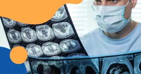
Multiple sclerosis (MS) doesn’t only show up on brain scans. Your neurologist also pays close attention to your cervical spine, the part of your backbone that runs through your neck. A magnetic resonance imaging (MRI) scan of the cervical spine can look very different depending on whether MS lesions are present, and those differences carry real weight in an MS diagnosis.
Here we’ll discuss what cervical spine lesions from MS look like compared with a normal scan, and when your doctor might order a cervical spine MRI.
Doctors often review a type of scan called a T2-weighted image, which shows areas of damage or inflammation as bright white spots. In a healthy scan, you won’t see these bright spots inside the cord itself.
Getting a clear picture isn’t always simple, though. The spinal cord is much smaller than the brain, and even slight movement can blur the image, so radiologists typically rely on higher-strength MRI machines to get a sharp, readable scan.
Many neurologists also compare cervical spine images with brain scans to get a more complete picture of MS, since lesions can occur throughout the central nervous system (CNS), which includes the brain and spinal cord. MS brain lesions also have specific features that help distinguish them from a healthy brain scan.
MS lesions on a cervical spine MRI have a few telltale features that set them apart from a healthy scan, and even from lesions caused by other conditions.
MS lesions in the spinal cord tend to be small. They usually don’t stretch across more than two vertebrae in length, and they don’t take up the full width of the cord. That’s different from some other conditions, where spinal cord lesions tend to be longer, larger, and can even cause the cord to swell.
MS lesions also tend to form along the sides or back portions of the spinal cord’s white matter (the area containing myelin-coated nerve fibers). The cervical section is a common location for these lesions, which is one reason doctors scan this area closely when checking for MS-related spinal cord lesions.
Doctors can often tell the difference between older lesions and ones that are actively inflamed. During the scan, they may use a contrast dye called gadolinium that lights up areas of fresh inflammation, which helps show which lesions are new versus older ones that have settled down.
Finding a mix of older and newer lesions in different parts of your spine and brain over time counts toward the criteria doctors use to diagnose MS.
If your MRI shows a new or larger lesion in your cervical spine, it’s information your care team will use to guide your treatment plan.
Spinal cord lesions carry real weight when it comes to your long-term outlook. Most people are diagnosed with relapsing-remitting MS, the most common form, marked by flare-ups followed by periods of recovery.
Even one lesion in the cervical spine found before age 30 has been linked to a higher chance of more severe disability and a faster shift toward a more progressive form of MS.
That doesn’t mean a new lesion is a reason to panic, but it is exactly the kind of finding your neurologist will factor in when deciding whether to start or switch to a different disease-modifying therapy (DMT). New or growing lesions can be one of the clearest signs that MS is still active, even if you haven’t noticed any new symptoms yourself.
Your neurologist may recommend a cervical spine MRI at a few different points along your MS journey.
When doctors are first working through a possible MS diagnosis, they typically order a cervical spine MRI along with a brain MRI. This combination helps confirm the diagnosis, rules out other causes of your symptoms, and creates a baseline picture of how much of your central nervous system has been affected.
Cervical spine imaging can even pick up signs of MS when a brain MRI comes back unclear or negative. Spinal cord lesions are also one of the specific locations doctors check when applying the McDonald criteria used to diagnose MS.
In one study, adding a cervical spine scan to a brain MRI raised the rate of people who met one of the official diagnostic criteria from about 78 percent to over 90 percent. This is just one example of how MRI supports an MS diagnosis.
Your doctor might also order a cervical spine MRI if you develop new symptoms that seem to come from your spinal cord, such as numbness, weakness, or the electric-shock feeling down your back known as Lhermitte’s sign. Lhermitte’s sign affects roughly 1 in 3 people with MS at some point.
Unlike brain MRIs, cervical spine scans aren’t usually repeated on a strict schedule once you’re diagnosed, since spinal cord symptoms tend to be noticeable on their own. Still, if your current treatment doesn’t seem to be working as well as expected, your neurologist may order a repeat scan to check for new activity and reassess your prognosis.
If you’re starting a new DMT, your care team will likely want periodic brain MRIs, roughly every six to 12 months, to see whether treatment is working. Spine follow-ups are used more selectively, since repeat imaging of that area can be technically harder to interpret.
 What MyMSTeam Members Say About Their Cervical Spine MRI
MyMSTeam members often discuss how cervical spine MRIs help explain symptoms that don’t always show up on a brain MRI. One member shared that although their brain MRI had been stable since 2011, a cervical spine MRI revealed a “significant interval increase” in lesions after months of worsening balance and walking “like I’m drunk.”
Another described a “new active lesion” in the cervical spine after developing bowel changes, hip pain, and other symptoms, leading their neurologist to recommend changing DMTs.
Some members also offer reassurance about the scan itself. One wrote that the MRI was “not nearly as difficult as anticipated,” thanks to headphones, a pillow, a warm blanket, and time to get comfortable.
Others remind members that symptoms such as arm or hand pain aren’t always caused by spinal cord lesions and encourage talking with a neurologist and getting a cervical spine MRI when appropriate.
On MyMSTeam, people share their experiences with multiple sclerosis, get advice, and find support from others who understand.
Have you had a cervical spine MRI as part of your MS diagnosis or ongoing care? Let others know in the comments below.
Get updates directly to your inbox.


How do spinal lesions affect prognosis



 Continue with Facebook
Continue with Email
Continue with Facebook
Continue with Email
Continue with Facebook
Continue with Email
Continue with Facebook
Continue with Email
Become a member to get even more





 Join
Join


 Your Privacy Choices

This is a member-feature!
Sign up for free to view article comments.
Let's you know the current state-of-the-art in diagnosis.
We'd love to hear from you! Please share your name and email to post and read comments.
You'll also get the latest articles directly to your inbox.